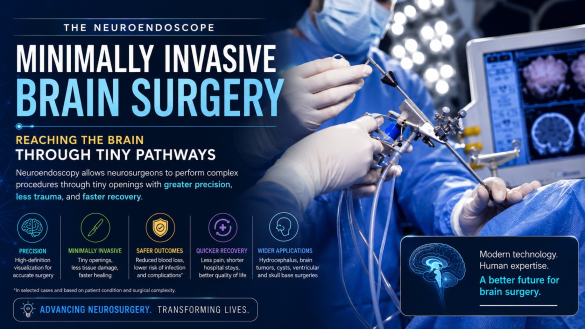
Reaching the Brain Through Tiny Pathways
In the past, complex brain procedures were performed by neurosurgeons using large incisions to access the brain that resulted in long recovery periods and increased complication rates. Advances in imaging and minimally invasive surgery have completely changed this procedure.
The development of the neuroendoscope provides neurosurgeons with the ability to perform surgeries using very small openings and with incredible accuracy. Neuroendoscopic surgery has revolutionized modern neurosurgery by decreasing the surgical trauma while also providing greater accuracy, safety, and recovery for the patient.
The Idea Behind Neuroendoscopy
Beginning with one major question, ‘How can I look inside of the human body/through to its interior without having to make a big incision?’ through to the instruments used in the 19th century to visualize the inside of hollow organs using bronze tubes and a glass mirror with a light bulb illuminated by a battery power source was the beginning of what we now regard as ‘neuroendoscopy.’ The application of this technology to the depths of the brain where the skull is very rigid and the tissues and structures are extremely delicate and sensitive created a huge challenge for many years before surgeons were able to safely penetrate/enter these areas of the human brain through small incision sites in the skull or elsewhere on head and/or face.
The Birth of Neuroendoscopy
The development of neuroendoscopy began in the early 1900s, as neurosurgeons were looking for a safer way to access difficult parts of the brain. One of the early pioneers was Walter Dandy, who developed surgical approaches for accessing the fluid-filled spaces (ventricles) in the brain.
Neuroendoscopy was formerly performed with instruments that did not provide adequate light or visibility. With advances in fiber optics, miniature cameras, high definition imaging systems and microsurgical instrumentation, neuroendoscopy has become an effective method of conducting surgery.
How Fiber Optics Revolutionized Brain Surgery
The use of fiber optics not only has numerous other benefits, but it has also transformed many aspects of surgical practice. With these tools now available, neurosurgeons can use them to more accurately visualize the internal structures of the brain than could ever have been done with conventional methods of performing surgery. Due to their properties as a conduit for light transmission, fibers enable neurosurgeons to see parts of the brain that they previously could only imagine being able to see. Because of this new capability, it is now possible for many procedures that used to be done with large incisions to be performed using small instruments and with direct visualization through real-time imaging, allowing for the introduction of modern neuroendoscopic techniques.
What Is a Neuroendoscope?
Neuroendoscope is a small, long instrument resembling a pen that enables neurosurgeons to look at the brain and carry out surgery through minimal incision. Each neuroendoscope is equipped with an HD camera, fibre optic light source, microsurgical instruments to guide the surgeon through each channel, an irrigation tube and a suction tube.
The real-time image captured by the camera will be sent to an external monitor so that neurosurgeons can conduct very accurate operations on an area of the body without damaging other parts. Depending on the surgical needs, neuroendoscopes can have rigid or flexible designs.
How Neuroendoscopic Surgery Is Performed
Neuroendoscopic surgery is performed with a burr hole (with the help of a neuroendoscope) into the brain. As the neuroendoscope has a live video feed, the surgeon is able to reach the desired area through as minimally invasive method as possible with very little harm to the surrounding tissue in the brain.
Once the surgeon completes the neuroendoscopic surgery, several specialized surgical instruments can be used through the neuroendoscope to perform any of the following: tumor/cyst removal, biopsy, drainage of cerebrospinal fluid, treatment of hydrocephalus and cessation of bleeding.
As neuroendoscopic surgery is much less invasive than traditional forms of brain surgery, the stress to the patient’s body is also much less versus if they had traditional open brain surgery performed.
Major Applications of Neuroendoscopy
- Hydrocephalus Treatment: Endoscopic Third Ventriculostomy (ETV) restores cerebrospinal fluid flow and reduces brain pressure.
- Brain Tumor Surgery: Used to remove certain tumors with minimal damage to surrounding tissue.
- Brain Cyst Removal: Allows minimally invasive drainage or removal of cysts inside the brain.
- Ventricular and Skull Base Procedures: Used in surgeries involving the brain ventricles and skull base, sometimes through the nasal cavity.
Advantages of Minimally Invasive Brain Surgery
Compared to traditional open brain surgery, neuroendoscopy offers several benefits:
- Smaller incisions with less tissue damage
- Reduced blood loss during surgery
- Faster recovery and shorter hospital stays
- Less postoperative pain
- Lower risk of infection and surgical complications in selected cases
Challenges and Limitations
There are many advantages of using neuroendoscopy in treating brain tumors; however, in cases where the size or vascularity (amount of blood vessel) of the tumor is too great, open surgery will likely be required instead of neuroendoscopic techniques. Because of their complexity and delicate nature, neuroendoscopic procedures require highly specialized skills and specialized equipment. The locations of many of the major brain pathways are very small, meaning that neuroendoscopic procedures are highly reliant on precision, accuracy, and surgical expertise.
The Future of Neuroendoscopy
The future of neuroendoscopy is being shaped by improvements in robotics, imaging, and AI technologies.
Examples of the new technologies being developed include robotic neuroendoscopes, 3D imaging systems, AI-enhanced surgical navigation systems, and improved flexible endoscopes. In addition, there are now better and more accurate methods of performing real-time image analysis on patients during their procedures.
The purpose of these new technologies is straightforward: to provide safer, more precise surgical techniques for your brain surgery with fewer deformities caused by your operation.
Conclusion
A neuroendoscope is a tool used to perform brain surgeries that provides surgeons with greater safety and precision than ever before by allowing them access to the brain through small openings. This represents an incredible new tool in the field of modern neurosurgery.
Thanks to advances in imaging and minimally invasive techniques, neurosurgeons now have a greater ability to accurately complete procedures, leading to less trauma and quicker recovery times for those patients undergoing treatment for neurosurgery.